38 chlamydomonas diagram with labels
Draw a neat labelled diagram. Chlamydomonas - Shaalaa.com Draw a neat labelled diagram. Chlamydomonas . Maharashtra State Board HSC Science (General) 11th. Textbook Solutions 8018. Important Solutions 19. Question Bank Solutions 5546. Concept Notes & Videos 432. Syllabus. Advertisement Remove all ads. Draw a neat labelled diagram. ... Chlamydomonas - Meaning, Structure, Life Cycle, Function and FAQs - VEDANTU Every flagellum has two contractile vacuoles at the base. A small red eyespot can be found on the chloroplast's anterior side. Given below is the Chlamydomonas structure with labels. The Life Cycle of Chlamydomonas . Chlamydomonas Reproduction is both sexual as well as asexual reproduction. Asexual reproduction takes place by following methods: 1.
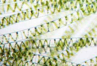
Spirogyra Labelled Diagram Spirogyra (common names include water silk, mermaid's tresses, and blanket weed) is a genus of filamentous charophyte green algae of the order Zygnematales, named for the helical or spiral arrangement of the chloroplasts that is characteristic of the genus. Draw a labelled diagram of Spirogyra. 51 Differentiate between flying lizard and bird.

Chlamydomonas diagram with labels
Labeled Diagram of Spirogyra - QS Study Labeled Diagram of Spirogyra. Plant kingdom. Spirogyra is a sophisticated, filamentous green alga, found in freshwater represented by about 300 species. It is also identified as pond silk, as its fiber burnishes like silk due to the occurrence of mucilage. Amoeba Diagram Illustrations, Royalty-Free Vector Graphics ... - iStock Browse 65 amoeba diagram stock illustrations and vector graphics available royalty-free, or start a new search to explore more great stock images and vector art. Newest results. Anatomy of an amoeba. Amoeba unicellular animal with pseudopods that lives in fresh or saltwater. Anatomy of an amoeba. Use this labeled diagram of a chlamydomonas cell to Use this labeled diagram of a Chlamydomonas cell to address the following two questions. 32. Which of the following statements correctly identifies aspects related to photosynthesis and/or respiration? 1. Acetyl CoA is most often found in G. 2. NADPH accumulates in C. 3. ATP is found in F. 4.
Chlamydomonas diagram with labels. Chlamydomonas | Facts, Structure, Life Cycle, & Classification Chlamydomonas, genus of biflagellated single-celled green algae (family Chlamydomonadaceae) found in soil, ponds, and ditches. Chlamydomonas species can become so abundant as to colour fresh water green, and one species, C. nivalis, contains a red pigment known as hematochrome, which sometimes imparts a red colour to melting snow. The cells of most Chlamydomonas species are more or less oval ... Animal Cells: Labelled Diagram, Definitions, and Structure Only present in lower plant forms (e.g. chlamydomonas) Present in all animal cells: Chloroplast: Plant cells have chloroplasts to synthesize their own food. Absent: Plasma Membrane: Cell wall and a cell membrane: Only cell membrane: Flagella: Present in some cells (e.g. sperm of bryophytes and pteridophytes, cycads and Ginkgo) Structure of Chlamydomonas (With Diagram) | Genetics In this article we will discuss about the structure of chlamydomonas (explained with labelled diagram). The unicellular green alga Chlamydomonas is haploid with a single nucleus, a chloroplast and several mitochondria (Fig. 9.3). It can reproduce asexually as well as sexually by fusion of gametes of opposite mating types (mt + and mt - ). Describe the structure of chlamydomonas with neat labelled diagram ... answeredOct 30, 2020by Naaji(56.8kpoints) selectedOct 30, 2020by Jaini Best answer 1. Chlamydomonas is a simple, unicellular, motile fresh water algae. They are oval, spherical or pyriform in shape. 2. The cell is surrounded by a thin and firm cell wall made of cellulose. 3. The cytoplasm is seen in between the cell membrane and the chloroplast. 4.
Structure and Diagram of Volvox and Their Functions Volvox Structure: Diagram of Volvox with Label The cells of anterior end possess bigger eye spots than those of posterior end cells. The cells of posterior side become reproductive on maturity. Thus, spherical or round colony of Volvox shows clear polarity. Cell structure of volvox colony are Chlamydomonas type. Chlamydomonas: Position, Occurrence and Structure (With Diagrams) Chlamydomonas is unicellular, motile green algae. The thallus is represented by a single cell. It is about 20 p,-30|i in length and 20 µ in diameter. The shape of thallus can be oval, spherical, oblong, ellipsoidal or pyriform. The pyriform or pear shaped thalli are common, they have narrow anterior end and a broad posterior end (Fig. 1). Life Cycle of Chlamydomonas (With Diagram) - Biology Discussion Each daughter cell develops cell wall, flagella and transforms into zoospore (Fig. 6). The zoospores are liberated from the parent cell or zoosporangium by gelatinization or rupture of the cell wall. The zoospores are identical to the parent cell in structure but smaller in size. The zoospores simply enlarge to become mature Chlamydomonas. Chlamydomonas - Wikipedia Drawings of Chlamydomonas caudata Wille. [1] Cross section of a Chlamydomonas reinhardtii cell Light micrograph of Chlamydomonas with two flagella just visible at bottom left Chlamydomonas globosa, again with two flagella just visible at bottom left
Solved: Label this diagram of the Chlamydomonas life cycle. | Chegg.com LearnSmart Online for Biology (10th Edition) Edit edition. This problem has been solved: Solutions for Chapter 21 Problem 24TY: Label this. diagram of the. Chlamydomonas life cycle.…. Get solutions. Get solutions Get solutions done loading. Looking for the textbook? How to make label Diagram of chlamydomonas - YouTube watch: "How to make thumbnail our you tube videos Hindi /urdu haris by #Top2utv" ... LABORATORY 9 - Susquehanna University Labeled diagram of Chlamydomonas. ... Chlamydomonas from culture. Cells have been stained with Lugol's Iodine, which complexes with true starch to turn black. 400X . You have slides of colonial volvocine green algae, which include Volvox, Gonium , Eudorina, ... Genetic map of the Chlamydomonas reinhardtii plastid genome ... Download scientific diagram | Genetic map of the Chlamydomonas reinhardtii plastid genome. Protein-coding regions are yellow and their exons are labeled with an "E" followed by a number denoting ...
Chlamydomonas - Lexikon der Biologie
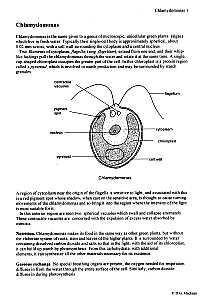
Biological drawings. Structure of Chlamydomonas. Learning Resources for ... Structure of Chlamydomonas: Next Drawing > Chlamydomonas is the name given to a genus of microscopic, unicellular green plants (algae) which live in fresh water. Typically their single-cell body is approximately spherical, about 0.02 mm across, with a cell wall surrounding the cytoplasm and a central nucleus.

Chlamydomonas study shows nature’s coping mechanism Algae Industry Magazine
Chloroplast Structure and Function in detail with Labelled Diagram The chloroplasts are the cell organelles which consist of these pigments. The 3 types of pigments present in plants are chlorophyll, carotenoids, and anthocyanins. Chlorophyll imparts the green color to plants. Plastids are membrane-bound cytoplasmic organelles that can be found in the cells of plants and algae.
Chlamydomonas Diagram With Labels
Solved: Chapter 21 Problem 24TY Solution - Chegg ISBN-13: 9780077388508 ISBN: 007738850X Authors: Sylvia S Mader Rent | Buy. This is an alternate ISBN. View the primary ISBN for: Biology 10th Edition Textbook Solutions.

The Chlamydomonas Genome Reveals the Evolution of Key Animal and Plant Functions | Science
Chlamydomonas as a Model Organism - Rice University Chlamydomonas as a Model Organism. Chlamydomonas, a genus of unicellular photosynthetic flagellates, is an important model for studies of such fundamental processes as photosynthesis, motility, responses to stimuli such as light, and cell-cell recognition.C. reinhardi, the most commonly studied species of Chlamydomonas, has a relatively simple genome, which has been sequenced.

DRAW IT NEAT : How to draw Chlamydomonas
Structure of Chlamydomonas (With Diagram) | Chlorophyta In this article we will discuss about the structure of chlamydomonas with the help of suitable diagrams. Chlamydomonas is unicellular, motile green algae. The thallus is represented by a single cell. It is about 20 p,-30|i in length and 20 µ in diameter. The shape of thallus can be oval, spherical, oblong, ellipsoidal or pyriform.

antibody labeling antibody 1 - Top Label Maker
Asymmetric properties of the Chlamydomonas reinhardtii cytoskeleton ... The C. reinhardtii eyespot. (a) A diagram illustrating asymmetric localization of the eyespot relative to the cytoskeleton. Two flagella and four microtubule rootlets extend from a pair of basal bodies at the anterior end of the cell; both the mother basal body (small black oval) and the daughter basal body (small gray oval) are associated with a four-membered rootlet (M4 or D4) and a two ...

Fig. 18-12. Schematic diagram of chlamydomonas
Morphology of Chlamydomonas (With Diagram) | Algae In this article we will discuss about the external morphology of chlamydomonas. Also learn about its Neuromotor Apparatus, Electron Micrograph, Palmella-Stage with suitable diagram. 1. The organism is an unicellular alga (Fig. 11). 2. The thallus is spherical to oblong in shape but some species are pyriform or ovoid. ADVERTISEMENTS: 3.

Bio Exam 1 Flashcards by ProProfs
Eye Diagram With Labels and detailed description - BYJUS A brief description of the eye along with a well-labelled diagram is given below for reference. Well-Labelled Diagram of Eye The anterior chamber of the eye is the space between the cornea and the iris and is filled with a lubricating fluid, aqueous humour. The vascular layer of the eye, known as the choroid contains the connective tissue.
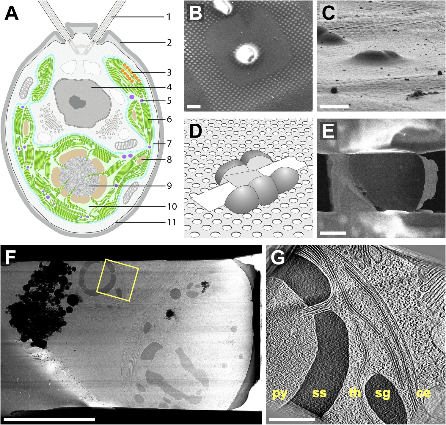
Native architecture of the Chlamydomonas chloroplast revealed by in situ cryo-electron ...
Diagram of Chlamydomonas angulosa... - Getty Images UNSPECIFIED - CIRCA 2003: Diagram of Chlamydomonas angulosa, Flagellated Protozoan. Drawing. (Photo by DeAgostini/Getty Images)

Downfield and upfield region of the 1D 1 H NMR spectra (600 MHz) at 298... | Download Scientific ...
Chlamydomonas reinhardtii - an overview | ScienceDirect Topics Chlamydomonas reinhardtii cells are oval shaped, c. 10 μm in length and 3 μm in width, with two flagellae at their anterior end (Figure 1). The cells contain a single chloroplast occupying 40% of the cell volume and several mitochondria. ... Diagram labeling densities in the averaged image. (B) Image average from thin sections of pf14 ...

AP Biology-Ch.6 A Tour of the Cell
Use this labeled diagram of a chlamydomonas cell to Use this labeled diagram of a Chlamydomonas cell to address the following two questions. 32. Which of the following statements correctly identifies aspects related to photosynthesis and/or respiration? 1. Acetyl CoA is most often found in G. 2. NADPH accumulates in C. 3. ATP is found in F. 4.
Chlamydomonas - Biology 1122
Amoeba Diagram Illustrations, Royalty-Free Vector Graphics ... - iStock Browse 65 amoeba diagram stock illustrations and vector graphics available royalty-free, or start a new search to explore more great stock images and vector art. Newest results. Anatomy of an amoeba. Amoeba unicellular animal with pseudopods that lives in fresh or saltwater. Anatomy of an amoeba.

Eyespot-Assembly Mutants in Chlamydomonas reinhardtii | Genetics
Labeled Diagram of Spirogyra - QS Study Labeled Diagram of Spirogyra. Plant kingdom. Spirogyra is a sophisticated, filamentous green alga, found in freshwater represented by about 300 species. It is also identified as pond silk, as its fiber burnishes like silk due to the occurrence of mucilage.

Chlamydomonas Diagram With Labels
Chlamydomonas Diagram With Labels

What is a Cell
Post a Comment for "38 chlamydomonas diagram with labels"